
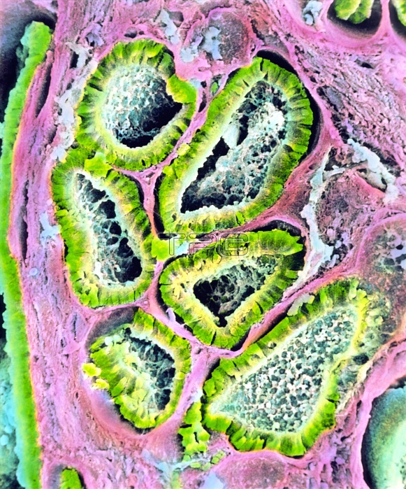
Sertoli-Leydig tumour in ovary. Coloured Scanning Electron Micrograph (SEM) of a section through a Sertoli-Leydig tumour in the human ovary. Known also as an arrhenoblastoma" or "androblastoma", up to 30% of these tumours are malignant (cancer- ous). This well differentiated tumour is made up of tubules or branching cords (green) lined with cuboidal or columnar Sertoli cells. Between the tubules is an interstitial component made up of Leydig cells (light blue); the stroma of the ovary is pink. A Sertoli-Leydig tumour can produce hormones, and may cause masculinization in women. Magnification: x350 at 6x7cm size. x460 at 4x5""
| px | px | dpi | = | cm | x | cm | = | MB |
Details
Creative#:
TOP10214078
Source:
達志影像
Authorization Type:
RM
Release Information:
須由TPG 完整授權
Model Release:
N/A
Property Release:
N/A
Right to Privacy:
No
Same folder images:
 Loading