
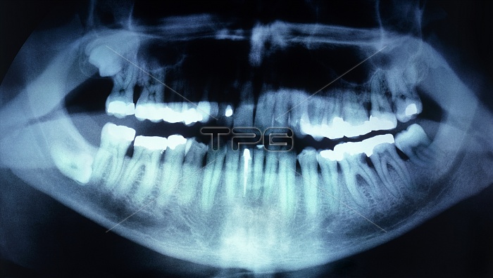
Dental fillings. X-ray of the mouth of a 45-year- old man. White areas show where repairs have been carried out on the teeth. The large white areas in the upper and lower molars (left and right) are fillings. Fillings are used to fill gaps caused by bacterial tooth decay. The rotten bit of the tooth is drilled out and a filling is inserted. The thin white area (lower centre) is where a dental cap (or crown) has been placed on one of the lower front teeth. It is held in place by a peg running down into the tooth. A dental cap is used to cover the visible surface of a tooth above the gumline. This panoramic wraparound X-ray clearly shows the twin roots of each molar.
| px | px | dpi | = | cm | x | cm | = | MB |
Details
Creative#:
TOP10212148
Source:
達志影像
Authorization Type:
RM
Release Information:
須由TPG 完整授權
Model Release:
N/A
Property Release:
N/A
Right to Privacy:
No
Same folder images:
 Loading