
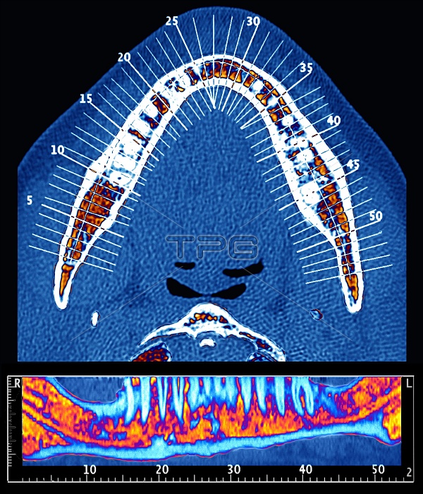
Dental surgery plan. Coloured computed tomography (CT) scans of the lower jaw of a patient, taken to plan surgery to implant dental prostheses. The two scans are in axial (horizontal, upper) and frontal (lower) aspects. The axial scan shows the arc of the jaw with its teeth (white). The frontal scan line (shown on the axial scan) runs parallel to the arc of the jaw. The teeth (blue) are embedded in the jaw (orange). In this case, the bone seemed to be too fragile to secure the dental implants. Also shown on the axial scan are the positions of 54 vertical scans taken at right-angles to the jaw arc. See image M780/335 for examples of these right-angle views.
| px | px | dpi | = | cm | x | cm | = | MB |
Details
Creative#:
TOP10211864
Source:
達志影像
Authorization Type:
RM
Release Information:
須由TPG 完整授權
Model Release:
N/A
Property Release:
N/A
Right to Privacy:
No
Same folder images:
 Loading