
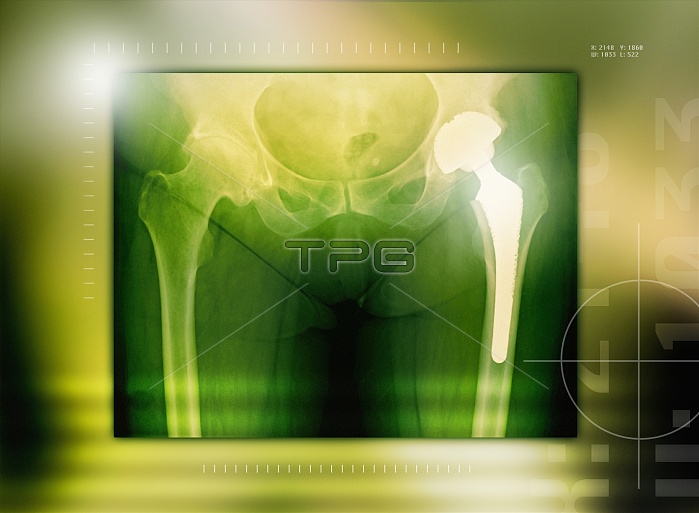
Hip replacement. Coloured X-ray of the pelvis of a female patient who has had a hip replacement. The prosthetic hip joint is on the right (white). It consists of a shaft and ball attached to the top of the femur (thigh bone) and a socket (bright semi-circle above the ball) that is inserted into the pelvis. The majority of hip replacements are a result of osteoarthritis, a degenerative joint disease that results in the loss of cartilage between the joint and the growth of bone in place of the cartilage. This causes pain, stiffness and loss of mobility.
| px | px | dpi | = | cm | x | cm | = | MB |
Details
Creative#:
TOP10208809
Source:
達志影像
Authorization Type:
RM
Release Information:
須由TPG 完整授權
Model Release:
N/A
Property Release:
N/A
Right to Privacy:
No
Same folder images:
 Loading