
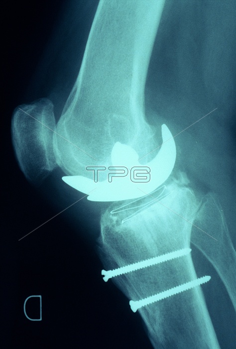
Prosthetic knee. X-ray of a side view of a knee joint with an artificial or prosthetic joint (bright area). The head of the femur of the upper leg has been replaced with an implant. The tibia, the larger of the two bones in the lower leg, has been fitted with two screws, probably to pin the bone together during healing of a fracture. The kneecap or patella is also seen (far left). Knee-joint replacement may be carried out following an accident to the leg, or it may be necessary in patients severely affected by osteoarthritis or rheumatoid arthritis, which cause inflammation and erosion of the joints.
| px | px | dpi | = | cm | x | cm | = | MB |
Details
Creative#:
TOP10208796
Source:
達志影像
Authorization Type:
RM
Release Information:
須由TPG 完整授權
Model Release:
N/A
Property Release:
N/A
Right to Privacy:
No
Same folder images:
 Loading