
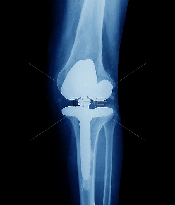
Knee replacement. Coloured X-ray of the prosthetic knee (white), seen from the front, of a patient with osteoarthritis. The implant attaches to the leg bones (blue/white), and has a flexible joint that can hinge like the old joint. The implant is attached to the top of the tibia (shin bone, lower frame) and to the bottom of the femur (thigh bone, upper frame). The other lower leg bone (fibula) is also seen (right of tibia). The implant replaced the old joint that had lost its cartilage due to osteoarthritis. Healthy cartilage reduces friction between the bones, and its progressive loss causes joint pain and immobility.
| px | px | dpi | = | cm | x | cm | = | MB |
Details
Creative#:
TOP10208743
Source:
達志影像
Authorization Type:
RM
Release Information:
須由TPG 完整授權
Model Release:
N/A
Property Release:
N/A
Right to Privacy:
No
Same folder images:
 Loading