
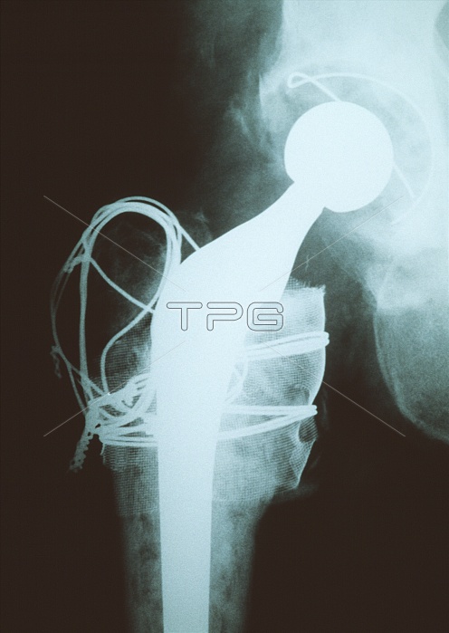
Hip replacement. X-ray showing a total artificial (prosthetic) replacement hip joint (white, upper right to bottom centre) in a 71-year-old man. The replacement consists of a ball and shaft, attached to the top of the femur (thigh bone, pale white, lower to bottom centre), and a socket, which is fitted into the pelvis (upper right). The ball is inserted into the socket. Surrounding the upper part of the shaft is a metal cage held in place by wires. Small pieces of bone (not seen) from the femoral head are placed in the cage to help graft the shaft to the bone. Hip replacements are most commonly carried out on older patients whose hips are painful as a result of arthritis.
| px | px | dpi | = | cm | x | cm | = | MB |
Details
Creative#:
TOP10208734
Source:
達志影像
Authorization Type:
RM
Release Information:
須由TPG 完整授權
Model Release:
N/A
Property Release:
N/A
Right to Privacy:
No
Same folder images:
 Loading