
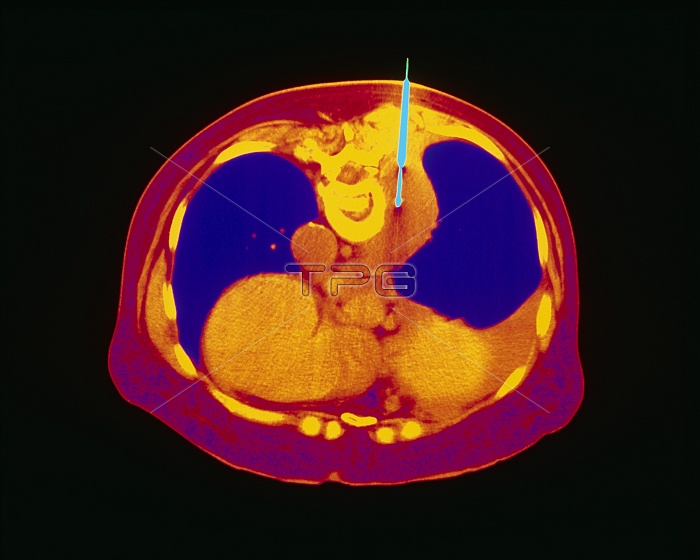
Lung biopsy. Coloured Computed Tomography (CT) scan of an axial section through the human chest, showing a lung biopsy being taken by brochoscope. The front of the chest is at bottom. Lung fields are coloured purple; the heart is at lower centre (orange). The bronchoscope (blue, at top) has penetrated the back of the patient next to the vertebral bone (yellow). It has entered a tumour (orange) which has formed on the lung. At the tip of the bronchoscope is a thin biopsy needle which extends to remove a tissue sample. The tissue is then examined for cell abnormalities such as found in cancer. The procedure using a needle such as this is also known as aspiration biopsy.
| px | px | dpi | = | cm | x | cm | = | MB |
Details
Creative#:
TOP10205454
Source:
達志影像
Authorization Type:
RM
Release Information:
須由TPG 完整授權
Model Release:
N/A
Property Release:
N/A
Right to Privacy:
No
Same folder images:
 Loading