
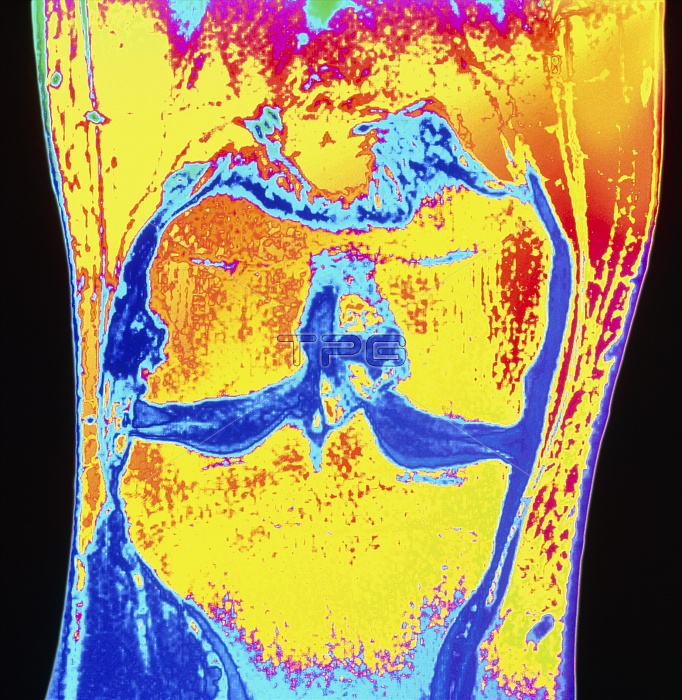
Knee injury. Coloured Magnetic Resonance Image (MRI) scan of the knee joint of a 35 year old man, showing meniscus degeneration. The head of the femur (thigh bone, upper centre) articulates with the tibia (shin bone, lower centre) at a central dark area. This dark region consists of cartilage discs called menisci which provide a smooth, strong surface for movement. Due to compression of the medial meniscus cartilage, the tibia bone is damaged. This appears as a blue line in the bone near the joint surface (centre right). It is due to natural wear and tear of the joint, exacerbated by the patient's high activity in sport.
| px | px | dpi | = | cm | x | cm | = | MB |
Details
Creative#:
TOP10202172
Source:
達志影像
Authorization Type:
RM
Release Information:
須由TPG 完整授權
Model Release:
N/A
Property Release:
N/A
Right to Privacy:
No
Same folder images:
 Loading