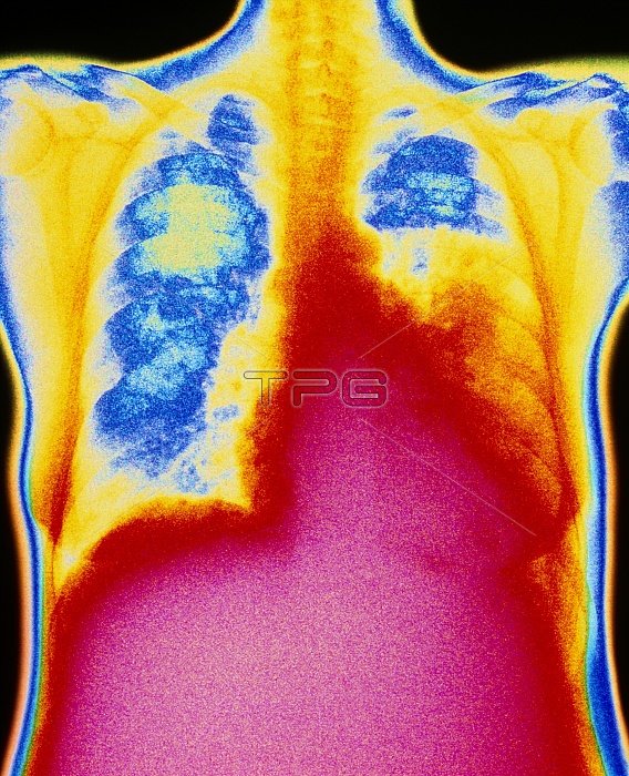
Lobar pneumonia. Coloured computer-enhanced chest X-ray showing lobar pneumonia in a patient's lung. The affected area of congestion appears in the lower lobe (orange) of the lung at right, above the pink heart. Normal lung tissue is blue/white. Bones of the neck and shoulders are yellow. Lobar pneumonia is caused by Streptococcus pneumoniae bacteria and initially affects a single lobe of one of the lungs. Alveoli (air sacs) in the lobe become blocked with pus, preventing air flow, causing tissue to solidify. Symptoms include fever, painful breathing and a cough that produces rust-coloured sputum. In most cases antibiotic drugs help the patient to make a full recovery.
| px | px | dpi | = | cm | x | cm | = | MB |
Details
Creative#:
TOP10200462
Source:
達志影像
Authorization Type:
RM
Release Information:
須由TPG 完整授權
Model Release:
N/A
Property Release:
N/A
Right to Privacy:
No
Same folder images:

 Loading