
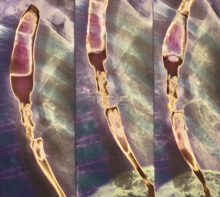
Constricted oesophagus. Coloured X-ray of a substance (brown) passing through a stricture in the oesophagus (gullet). The X-rays are sequenced from left to right. The stricture is seen at lower left, centre and centre right. The stricture may result from a tumour, inflammation (oesophagitis), or swallowing corrosive substances. The stricture blocks the normal passage from mouth to stomach, which results in difficulty swallowing and pain. Once cancer has been ruled out, treatment involves oesophageal dilation. If very severe or if a long segment is affected, the area may need to be surgically removed and replaced with a loop of the patient's colon (large intestine).
| px | px | dpi | = | cm | x | cm | = | MB |
Details
Creative#:
TOP10200284
Source:
達志影像
Authorization Type:
RM
Release Information:
須由TPG 完整授權
Model Release:
N/A
Property Release:
N/A
Right to Privacy:
No
Same folder images:
 Loading