
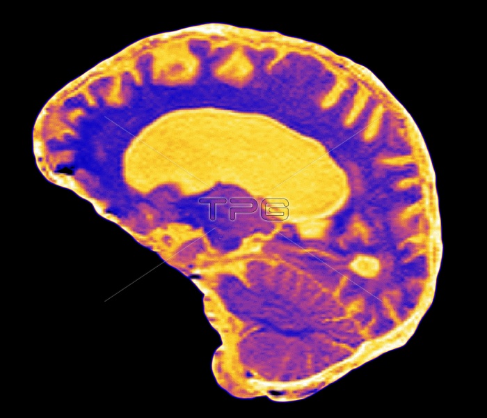
Multiple sclerosis. Coloured magnetic resonance imaging (MRI) scan of a sagittal (vertical) secti- on through a brain in multiple sclerosis (MS). The front of the brain is at left. In MS the myelin sheaths around the axon nerve fibres of the brain (purple) are destroyed. This produces demyelinated lesions such as the small yellow area at lower right, and abnormally enlarged fluid-filled ventr- icles (one at centre, yellow). Affected axons can no longer conduct nerve impulses, causing highly individual symptoms ranging from tingling to para- lysis. MS is thought to be an autoimmune disorder in which the immune system attacks myelin.
| px | px | dpi | = | cm | x | cm | = | MB |
Details
Creative#:
TOP10199922
Source:
達志影像
Authorization Type:
RM
Release Information:
須由TPG 完整授權
Model Release:
N/A
Property Release:
N/A
Right to Privacy:
No
Same folder images:
 Loading