
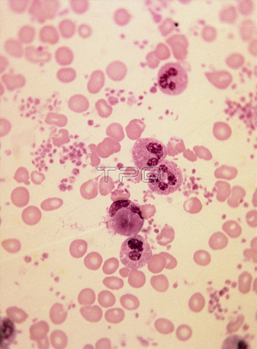
Light micrograph of a human blood smear, showing the characteristic appearance of white blood cells in lupus erythematosus (LE). In this smear, the second white cell from the top of field, a neutrophil, has phagocytosed (engulfed) the dark- pink staining nucleus of an LE cell. Other white cells, probably neutrophils, are visible in the smear, as are smaller, pink-staining red blood cells (disc-like) and even smaller platelets. LE is a multisystem disease, with a variety of immunological abnormalities involved in tissue damage. Magnification: x200 at 35mm size. Mag: x370 at 3x3-inch original size.
| px | px | dpi | = | cm | x | cm | = | MB |
Details
Creative#:
TOP10199724
Source:
達志影像
Authorization Type:
RM
Release Information:
須由TPG 完整授權
Model Release:
N/A
Property Release:
N/A
Right to Privacy:
No
Same folder images:
 Loading