
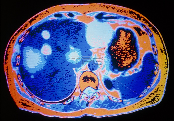
Hydatid disease. Coloured computed tomography (CT) scan showing cysts (light areas) in the liver (blue, left) due to hydatid disease. This is a horizontal slice through the abdomen, with the patient's back at the bottom of the picture. At lower centre is the spine (pale blue), at right the spleen (blue) and stomach (orange). Hydatid disease is caused by larvae of the tapeworm Echinococcus granulosus. Infection usually occurs in childhood, with the larvae settling in the liver (and sometimes the lungs, muscles or brain), and causing the development of slow-growing cysts. Symptoms, if any, appear in adults. They include tender lumps, bile duct obstruction and jaundice.
| px | px | dpi | = | cm | x | cm | = | MB |
Details
Creative#:
TOP10198850
Source:
達志影像
Authorization Type:
RM
Release Information:
須由TPG 完整授權
Model Release:
N/A
Property Release:
N/A
Right to Privacy:
No
Same folder images:
 Loading