
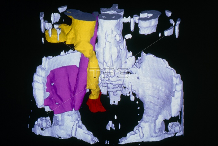
False-colour three-dimensional computed tomography (CT) scan of the pelvic region, showing a kidney and ureter diseased with hydronephrosis. Bone is coloured white. The base of the right kidney is seen (top left, yellow) greatly distended with urine, due to an obstruction in the urinary tract, particularly the ureter. Here, the ureter has swelled along its tube length, to the point (red) at which it joins the bladder. The retaining of fluid caused by hydronephrosis can cause irrepair- able damage to the kidneys. In this image the pelvic psoas muscle (violet) is also seen, arising from thoracic vertebrae; it flexes the trunk.
| px | px | dpi | = | cm | x | cm | = | MB |
Details
Creative#:
TOP10198833
Source:
達志影像
Authorization Type:
RM
Release Information:
須由TPG 完整授權
Model Release:
N/A
Property Release:
N/A
Right to Privacy:
No
Same folder images:
 Loading