
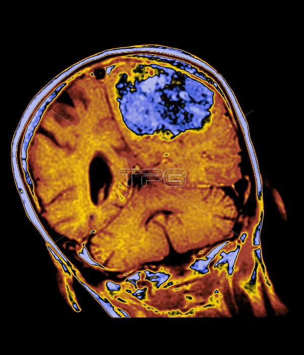
Brain tumour. Coloured magnetic resonance imaging (MRI) scan of a coronal section through the brain of a 74 year old woman, showing a large tumour. At upper centre is the tumour (blue) within one cerebral hemisphere (orange) of the brain; the other cerebral hemisphere (at centre left) is normal containing a dark ventricle or cavity. The cerebellum of the brain is seen at lower centre. Brain tumours may be primary tumours arising in the brain first or they may be spread from cancer elsewhere in the body. A large tumour such as this may cause brain compression and nerve damage.
| px | px | dpi | = | cm | x | cm | = | MB |
Details
Creative#:
TOP10197689
Source:
達志影像
Authorization Type:
RM
Release Information:
須由TPG 完整授權
Model Release:
N/A
Property Release:
N/A
Right to Privacy:
No
Same folder images:
 Loading