
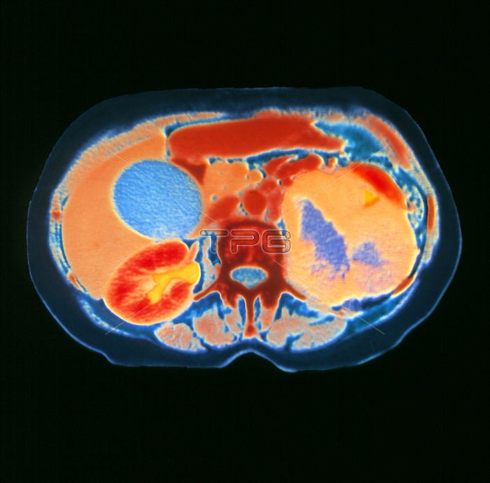
Kidney cancer. Coloured computed Tomography (CT) scan, axial section, of the human abdomen showing kidney cancer. At centre is a vertebra (rust). The diseased kidney (right on image, orange) is distorted and enlarged by a cancer tumour. This diseased kidney with a blue coloured interior is non-functioning except for a small area excreting into the collecting system (yellow triangle, upper right). At lower left is a normal kidney (red, bean-shaped), that is excreting an X-ray contrast agent (yellow). The liver (brown, left) is next to the normal kidney, with the circular gall bladder (blue) covering the liver.
| px | px | dpi | = | cm | x | cm | = | MB |
Details
Creative#:
TOP10197608
Source:
達志影像
Authorization Type:
RM
Release Information:
須由TPG 完整授權
Model Release:
N/A
Property Release:
N/A
Right to Privacy:
No
Same folder images:
 Loading