
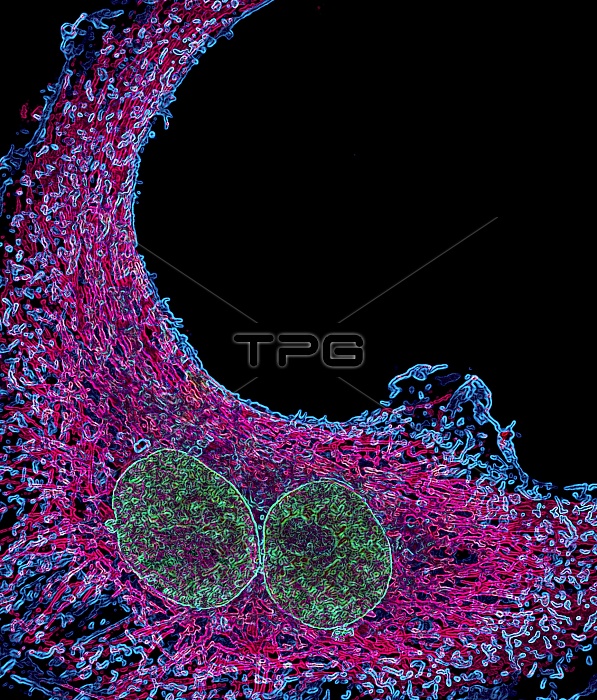
Cancer cell. Immunofluorescent light micrograph of a cancer cell containing two nuclei (binucleated). The distribution of DNA (green, in the nuclei), microtubules (pink) and actin (blue), is shown. Immunofluorescence uses antibodies to attach fluorescent dyes to specific cell tissues.
| px | px | dpi | = | cm | x | cm | = | MB |
Details
Creative#:
TOP10197362
Source:
達志影像
Authorization Type:
RM
Release Information:
須由TPG 完整授權
Model Release:
N/A
Property Release:
N/A
Right to Privacy:
No
Same folder images:
 Loading