
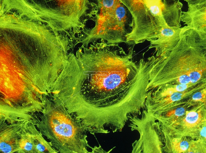
Immunofluorescent Light Micrograph of cancer cells of the human kidney, cultured in the laboratory. Such malignant cells typically are large and un- differentiated, with a capacity to divide rapidly. Here, the prominent nucleus of each cell stains blue; cytoplasm is orange/yellow. At lower left, numerous cells are undergoing mitotic division. Green fibres are actin stress fibres: strands of natural protein that form a support network around cultured cells. Immunofluorescence is a staining technique which uses antibodies to attach fluores- cent dyes to specific tissues and to molecules within the cell.Mag:x200 at35mm,x340 at 6x4.5size.
| px | px | dpi | = | cm | x | cm | = | MB |
Details
Creative#:
TOP10197076
Source:
達志影像
Authorization Type:
RM
Release Information:
須由TPG 完整授權
Model Release:
N/A
Property Release:
N/A
Right to Privacy:
No
Same folder images:
 Loading