
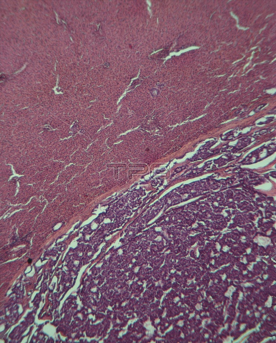
Liver cancer. Light micrograph of a metastatic carcinoma in the human liver. Here, normal liver cells (above centre) are seen in contrast with malignant tumour cells (purple, below centre). In undiseased liver tissue the cytoplasm (pink) is abundant, and nuclei are small (purple dots). The growing cancerous tissue has enlarged nuclei which are actively dividing, and cytoplasm is scant. A metastatic carcinoma is a secondary tumour that has spread from another primary site in the body: liver cancer may arise from cancer spread from the colon. There is no cure for secondary liver cancer but anticancer drugs slow the progress of this disease. Magnification: x40 at 6x7cm size.
| px | px | dpi | = | cm | x | cm | = | MB |
Details
Creative#:
TOP10197075
Source:
達志影像
Authorization Type:
RM
Release Information:
須由TPG 完整授權
Model Release:
N/A
Property Release:
N/A
Right to Privacy:
No
Same folder images:
 Loading