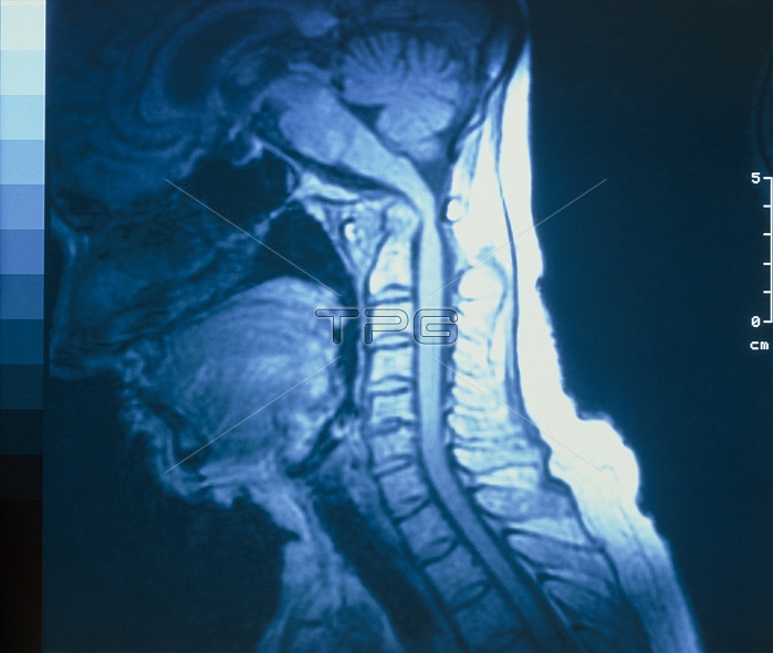
Rheumatoid arthritis. Magnetic resonance image (MRI) of the neck of a 70 years-old woman suffering from rheumatoid arthritis, showing the subluxation (partial dislocation) of the atlanto- axial joint. This joint between the top two cervical vertebrae (the atlas & axis) may become dangerously weakened in rheumatoid arthritis, due to destruction of the ligaments that bind the joint. As a result, the spinal cord has become compressed, and appears pinched & abnormally bent in this image. Symptoms of shooting pains & weakness in the limbs result; severe cases may produce quadriplegia & even sudden death.
| px | px | dpi | = | cm | x | cm | = | MB |
Details
Creative#:
TOP10195600
Source:
達志影像
Authorization Type:
RM
Release Information:
須由TPG 完整授權
Model Release:
N/A
Property Release:
N/A
Right to Privacy:
No
Same folder images:

 Loading