
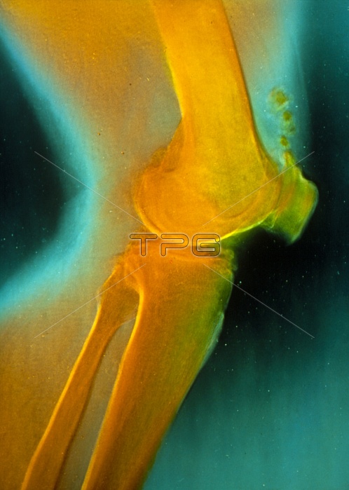
False-colour X-ray of a profile view of the human knee joint, showing osteoarthritic changes in the joint. The large bone at top is the femur (thigh bone), which articulates against the tibia (shin bone) and the fibula (bottom) at the knee. Immediately to the right of the head of the femur is the patella, the knee cap. In a normal, healthy joint the heads of the femur and tibia would not appear in direct contact, as they do in this image. Narrowing of the joint space due to loss of cartilage (the working surface of the joint) is a typical X-ray feature in osteoarthritis. The condition is associated with mechanical wear and is most common in persons over 50 years of age.
| px | px | dpi | = | cm | x | cm | = | MB |
Details
Creative#:
TOP10195586
Source:
達志影像
Authorization Type:
RM
Release Information:
須由TPG 完整授權
Model Release:
N/A
Property Release:
N/A
Right to Privacy:
No
Same folder images:
 Loading