
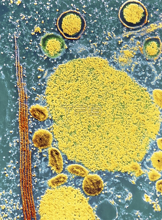
False-colour transmission electron micrograph (TEM) of a cell affected with the myxomatosis virus (a member of the poxvirus group), showing sites of viral synthesis & replication within the cell & viruses in different stages of evolution. The central yellow area can be considered as the site of viral synthesis & is known as the viroplasma. Viruses are seen in 3 stages of development. The larger circular particles at top (with separate surrounding membrane) are in 1st stage, the smaller, dark oval bodies at bottom left are in 2nd stage, & the nearby dark circular viruses are in the 3rd stage of development. Magnification: X 14,700 at 35mm size.
| px | px | dpi | = | cm | x | cm | = | MB |
Details
Creative#:
TOP10194298
Source:
達志影像
Authorization Type:
RM
Release Information:
須由TPG 完整授權
Model Release:
N/A
Property Release:
N/A
Right to Privacy:
No
Same folder images:
 Loading