
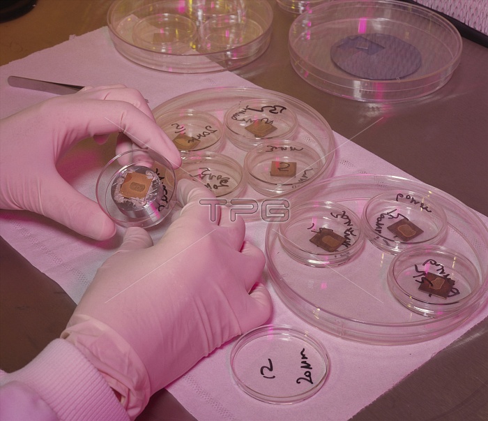
Spinal cord samples. Hands holding a section of human spinal cord sliced and mounted for scanning electron microscopy (SEM). The individual sections have been mounted on a thin copper plate and gold plated. This is to allow the non-conducting tissue a means of conducting the beam current of the microscope away. SEM involves sweeping a beam of electrons over the surface of the object to be studied, and collecting the electrons it gives off in response. It produces sharp 3-dimensional images and allows magnifications of up to 100,000 times.
| px | px | dpi | = | cm | x | cm | = | MB |
Details
Creative#:
TOP10193902
Source:
達志影像
Authorization Type:
RM
Release Information:
須由TPG 完整授權
Model Release:
N/A
Property Release:
N/A
Right to Privacy:
No
Same folder images:
 Loading