
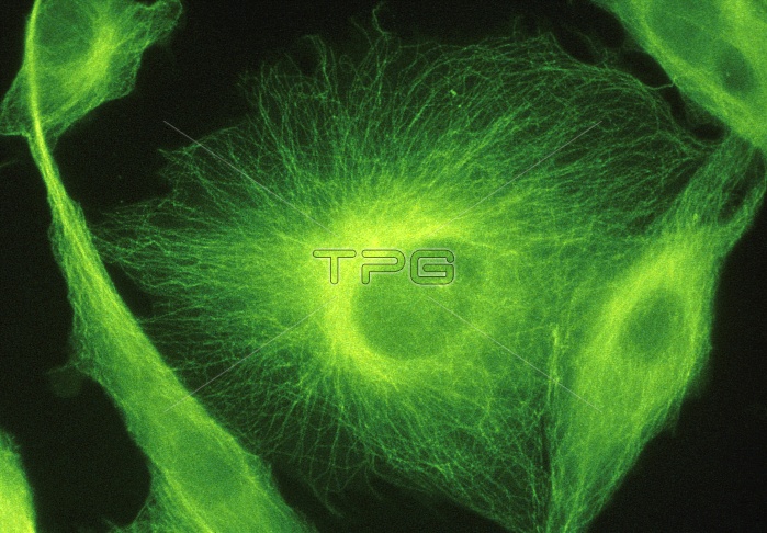
Ultraviolet fluorescence light micrograph of BHK (baby hampster kidney) tissue culture cells labelled with anti-tubulin to show the micro- tubular structure. Microtubules, together with microfilaments (not seen here), form a three- dimensional array known as the cytoskeleton. This fibrous network, only recently observed using ultraviolet light & fluorescent stains, is still poorly understood. Microtubules, more rigid than microfilaments, are thought to act as direction markers in the cell. In this image the oval depression at the centre of each cell is the space occupied by the nucleus. The microtubules radiate from the nucleus to the periphery of the cell.
| px | px | dpi | = | cm | x | cm | = | MB |
Details
Creative#:
TOP10188552
Source:
達志影像
Authorization Type:
RM
Release Information:
須由TPG 完整授權
Model Release:
N/A
Property Release:
N/A
Right to Privacy:
No
Same folder images:
 Loading