
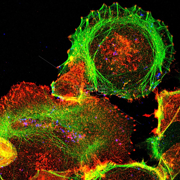
Cultured cell. Immunofluorescent light micrograph of an epithelial cell (round, upper right). The cell proteins are marked by fluorescent dyes. The actin cytoskeleton is green, bacteria are the blue dots, and red marks an activated protein called phosphotyrosine (a protein that plays a role in the regulation of cell growth). The actin fibres surrounding the cell at upper right end in areas of this activated protein. Yellow marks an overlap between green and red. Epithelial cells make up the body's internal and external surface layers, such as the skin and the walls of the intestines.
| px | px | dpi | = | cm | x | cm | = | MB |
Details
Creative#:
TOP10188279
Source:
達志影像
Authorization Type:
RM
Release Information:
須由TPG 完整授權
Model Release:
N/A
Property Release:
N/A
Right to Privacy:
No
Same folder images:
 Loading