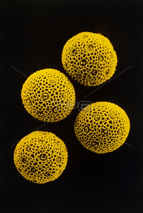
False co;our scanning electron micrograph showing four pollen grains from the flower of a Red Campion, Silene dioica.. These spherical pollen grains have many pores on their outer wall (multiporate) called germinal pores; each pore is capped by an operculum (lid) covered with wart- like processes (verra). Between the pores the surface appears as a network of ridges & irregular sized openings. When the pollen, containing the male gametes, lands on the stigma of the flower, it germinates & a pollen tube will grow out of one of the pores down to the ovary. The male nuclei travel down this tube, fertilize the ovules & a seed is formed. Magnification: X286 at 35mm size.
| px | px | dpi | = | cm | x | cm | = | MB |
Details
Creative#:
TOP10175216
Source:
達志影像
Authorization Type:
RM
Release Information:
須由TPG 完整授權
Model Release:
N/A
Property Release:
N/A
Right to Privacy:
No
Same folder images:

 Loading