
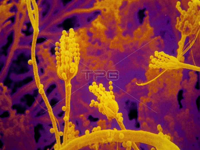
Fungus. Coloured scanning electron micrograph of a Penicillium sp. fungus. In this image, the condiophores are seen. They are the stalk-like objects, to which are attached numerous round condia. The conidia are the fruiting bodies of the fungus. Magnification: x900 at 6x4.5cm size.
| px | px | dpi | = | cm | x | cm | = | MB |
Details
Creative#:
TOP10167105
Source:
達志影像
Authorization Type:
RM
Release Information:
須由TPG 完整授權
Model Release:
N/A
Property Release:
N/A
Right to Privacy:
No
Same folder images:
 Loading