
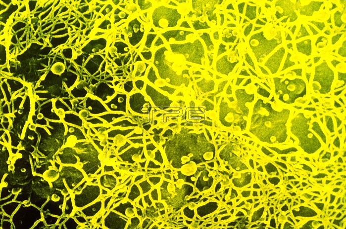
False-colour scanning electron micrograph of details of the surface of a tobacco leaf protoplast, Nicotiana tabacum (cv White Burley) showing the regrowth of the cell wall ten hours after culturing. The fibres visible correspond to cellulose microfibrils, beneath is the plasma membrane and this highlights the underlying chloroplasts. The spherical bodies visible among the fibres are either membrane blebs or artefacts. Magnification: x3430 at 35mm size.
| px | px | dpi | = | cm | x | cm | = | MB |
Details
Creative#:
TOP10166087
Source:
達志影像
Authorization Type:
RM
Release Information:
須由TPG 完整授權
Model Release:
N/A
Property Release:
N/A
Right to Privacy:
No
Same folder images:
 Loading