
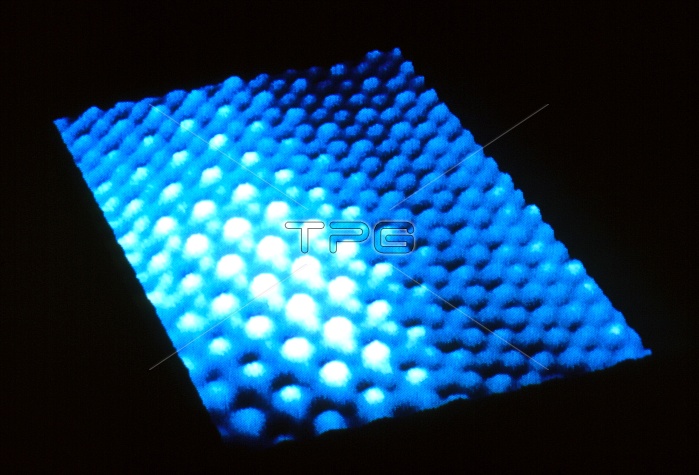
Scanning tunnelling micrograph of palladium atoms on a graphite substrate. The palladium atoms, shown as white, form a hexagonal arrangement with a spacing of 0. 426 nanometres. The graphite (carbon) atoms are shown as blue. The scanning tunnelling microscope (STM) uses a fine point electrode, a few atoms thick at the tip. This is brought to within a few angstroms of the sample's surface. As the electron clouds of the tip and the sample atoms interact, a small 'tunnelling' current passes. The electrode scans across the surface, moving vertically to maintain the tunnelling current. This motion is then processed to produce a map such as that seen here.
| px | px | dpi | = | cm | x | cm | = | MB |
Details
Creative#:
TOP10162728
Source:
達志影像
Authorization Type:
RM
Release Information:
須由TPG 完整授權
Model Release:
N/A
Property Release:
N/A
Right to Privacy:
No
Same folder images:
 Loading