
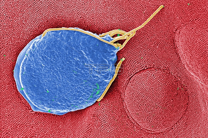
This digitally-colorized scanning electron micrograph (SEM) depicted a Giardia muris protozoan adhering itself to the microvillous border of an intestinal epithelial cell. Each small circular profile under the protozoan represents the rounded tip of a single microvillous, and it is estimated that 2000 to 3000 microvilli cover the surface of a single intestinal epithelial cell. The two circular lesions on the right side of the photograph are impressions made by the ventral adhesive disk of othe
| px | px | dpi | = | cm | x | cm | = | MB |
Details
Creative#:
TOP09810678
Source:
達志影像
Authorization Type:
RM
Release Information:
須由TPG 完整授權
Model Release:
No
Property Release:
No
Right to Privacy:
No
Same folder images:
 Loading