
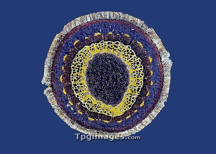
Virginia creeper stem. Polarised light micrograph of a cross-section through a stem from a Virginia creeper (Parthenocissus tricuspidatus). The primary epidermis has sloughed off and been replaced by an outer cork layer (white-brown). The next layer is the cortex (red and blue), which surrounds the vascular cylinder. The outer vascular layer is a broken ring of phloem (red, topped with yellow fibres). The next ring is xylem tissue (yellow). The pith (centre) is composed of large parenchyma cells (blue). Magnification: x14 when printed at 10 centimetres across.
| px | px | dpi | = | cm | x | cm | = | MB |
Details
Creative#:
TOP07421672
Source:
達志影像
Authorization Type:
RM
Release Information:
須由TPG 完整授權
Model Release:
NO
Property Release:
NO
Right to Privacy:
No
Same folder images:
virginiacreeperparthenocissustricuspidatustissuecellstemcreeperplantvinebiologybotanylightmicrographlmplmlightmicroscopecirclebiologicalcutoutsectionsectionedtransversecross-sectionpolarisedlightmicrographpolarizedpolarisedlightmicroscopycut-outcutoutcut-outscutoutscutoutscircularroundbotanicalnaturefloracellscellularxylemphloemvascularbundlevascularbundlesnutrienttransportwatertransport
biologicalbiologybotanicalbotanybundlebundlescellcellscellularcirclecircularcreepercreepercross-sectioncutcutcut-outcut-outscutoutcutoutsfloralightlightlightlightlmmicrographmicrographmicroscopemicroscopynaturenutrientoutoutsparthenocissusphloemplantplmpolarisedpolarisedpolarizedroundsectionsectionedstemtissuetransporttransporttransversetricuspidatusvascularvascularvinevirginiawaterxylem
 Loading