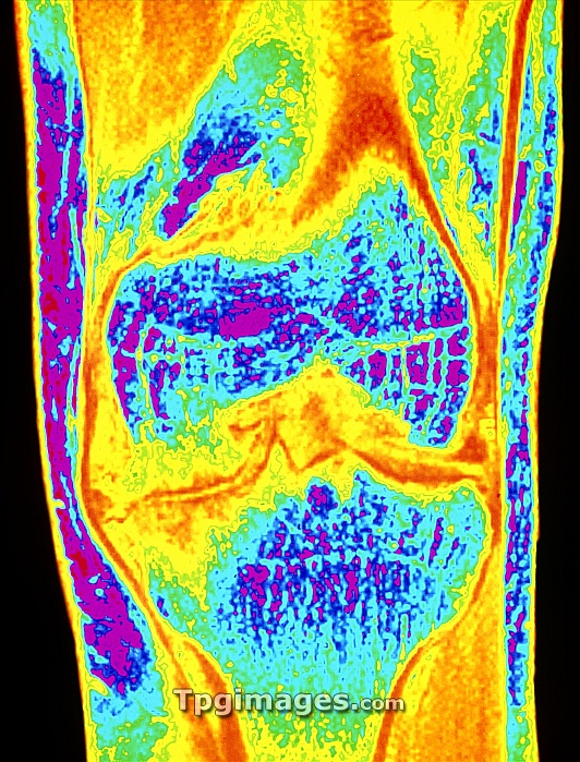
Osteoarthritis of knee. Colour Magnetic Resonance Imaging (MRI) scan of a front view of the knee joint of a 33 year old male with osteoarthritis. The lower end of the femur (thigh bone) is at centre (blue); it articulates with the upper end of the tibia (shin bone) at lower centre (blue). Due to osteoarthritis the ends of the bones have become severely eroded and jagged; cracking of the bone is also visible. A deep intercondylar notch (triangular) has formed in the femur. Osteo- arthritis is the most common type of arthritis due mainly to wear and tear on joints in the elderly. This patient had haemophilia which was a factor.
| px | px | dpi | = | cm | x | cm | = | MB |
Details
Creative#:
TOP06674049
Source:
達志影像
Authorization Type:
RM
Release Information:
須由TPG 完整授權
Model Release:
NO
Property Release:
NO
Right to Privacy:
No
Same folder images:

 Loading