
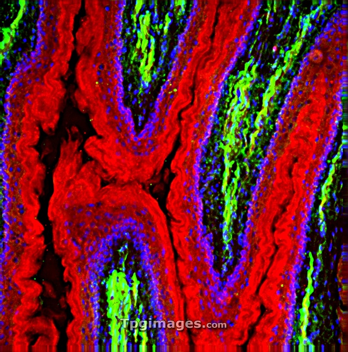
Oesophagus lining. Fluorescence deconvolution micrograph of a section through a healthy oesophagus (gullet), showing the muscle lining (g-actin, red). Green is the f-actin of muscle in the walls, and blue is cell nuclei. Magnification x40 when printed 10 centimetres wide.
| px | px | dpi | = | cm | x | cm | = | MB |
Details
Creative#:
TOP06663269
Source:
達志影像
Authorization Type:
RM
Release Information:
須由TPG 完整授權
Model Release:
NO
Property Release:
NO
Right to Privacy:
No
Same folder images:
humanhumanbodyanatomybiologyhistologymedicinelightmicrographfluorescentdeconvolutionmicrographconfocallightmicrographlightmicroscopeactinanatomicalbiologicalcellcellsesophagealesophagusfluorescenceg-actingastrointestinalgullethealthyhistologicalhistopathologicallmmedicalmusclesmuscularnormalnucleinucleusoesophagealoesophagusproteinsectionsectionedsmoothmusclestructuretissuewall"
"actinanatomicalanatomybiologicalbiologybodycellcellsconfocaldeconvolutionesophagealesophagusfluorescencefluorescentg-actingastrointestinalgullethealthyhistologicalhistologyhistopathologicalhumanhumanlightlightlightlmmedicalmedicinemicrographmicrographmicrographmicroscopemusclemusclesmuscularnormalnucleinucleusoesophagealoesophagusproteinsectionsectionedsmoothstructuretissuewall
 Loading