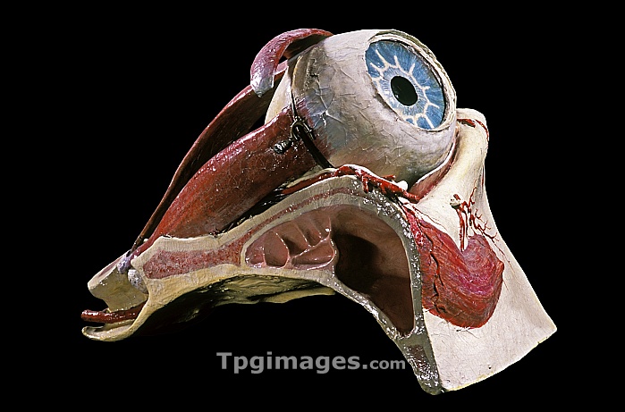
19th century anatomical model of an eye. This papier mache model was made to be used as a teaching aid by Louis Auzoux in about 1850. It is designed in different sections that fit together and can be separated to show the internal structure. Here the bone (white) structure of the eye socket and one of the paranasal sinuses (large space, centre) can be seen along with the eyeball (top) and some of the muscles (red) that control its movement. Auzoux was a pioneer of the construction of anatomical models. Photographed in the museum of the National Veterinary School of Alfort, Maisons-Alfort, France.
| px | px | dpi | = | cm | x | cm | = | MB |
Details
Creative#:
TOP06660368
Source:
達志影像
Authorization Type:
RM
Release Information:
須由TPG 完整授權
Model Release:
NO
Property Release:
NO
Right to Privacy:
No
Same folder images:

 Loading