
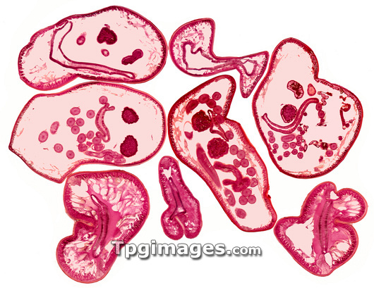
Roundworms. Light micrograph of cross-sections through various regions in several roundworms (Ascaris lumbricoides). The red edges are the cuticle and epidermis and the pale inner region is the pseudocoil. In the slice at upper centre, the gut can be seen inside the psuedocoel (body cavity), with a layer of epithelial cells (red) lining it. The central slice shows the worm's uteri (dark circles). At centre left, the small pink circles are egg cells (ova) in the ovary and at lower left is a slice showing muscle tissue (fringe of dark red fibres). The adult worm lives in the small intestine of vertebrates, where the female lays eggs. The eggs are excreted in the faeces and ingested by a new host through infected water or food. Magnification: x4 when printed 10cm wide.
| px | px | dpi | = | cm | x | cm | = | MB |
Details
Creative#:
TOP03229726
Source:
達志影像
Authorization Type:
RM
Release Information:
須由TPG 完整授權
Model Release:
N/A
Property Release:
N/A
Right to Privacy:
No
Same folder images:
 Loading