
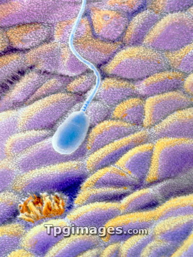
A spermatozoon on the uterine mucosa. False-colour scanning electron micrograph of a spermatozoon floating over the endometrium, the internal wall of the uterus. Spermatozoa have pear-shaped heads about 7 microns long and a tail about 60 microns long. This image also shows the two different types of simple columnar cells which populate this area: ciliated (orange bottom left) and secretory (pink, yellow). During their journey through the uterus towards the Fallopian tubes spermatozoa undergo a capacitation process which enable them to penetrate and then fertilise an egg. Magnification: x2200 at 6x7cm size. x3465 at 4x5ins
| px | px | dpi | = | cm | x | cm | = | MB |
Details
Creative#:
TOP03222011
Source:
達志影像
Authorization Type:
RM
Release Information:
須由TPG 完整授權
Model Release:
N/A
Property Release:
N/A
Right to Privacy:
No
Same folder images:
 Loading