
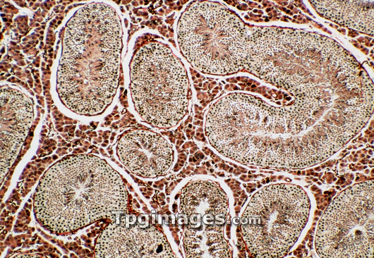
Light micrograph of a normal human testis, showing a cross section through the coiled system of seminiferous tubules. Each tubule, outlined by a continuous brown line, the basement membrane, holds a population of spermatogenic cells of differing maturity. Next to the membrane are the least mature spermatogonia, seen as black dots. They give rise to spermatocytes (brown dots), then spermatids, & finally spermatozoa (brown layer at tubule centre). Around the tubules lies the areolar tissue, source of testosterone (hormone). Black fibrous lines within this are blood vessel walls. Magnification: x25 at 35mm size.
| px | px | dpi | = | cm | x | cm | = | MB |
Details
Creative#:
TOP03221935
Source:
達志影像
Authorization Type:
RM
Release Information:
須由TPG 完整授權
Model Release:
N/A
Property Release:
N/A
Right to Privacy:
No
Same folder images:
 Loading