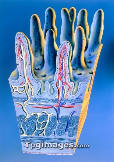
Illustration of intestinal villi. These are minute finger-like projections found on the lining of the small intestine. Their role is food absorption. Each villus contains a lymph vessel (white) and a network of blood capillaries (red and blue). Tall columnar cells on the surface epithelium absorb food that passes over the villi. This food in turn passes into the blood circulation. Found at the base of villi are indentations called Crypts of Lieberkuhn. These crypts produce the cells that go to make up each villus. At bottom can be seen other muscle layers of the intestinal wall. Villi are largest and most numerous in the duodenum and jejunum, where most food absorption occurs.
| px | px | dpi | = | cm | x | cm | = | MB |
Details
Creative#:
TOP03221751
Source:
達志影像
Authorization Type:
RM
Release Information:
須由TPG 完整授權
Model Release:
N/A
Property Release:
N/A
Right to Privacy:
No
Same folder images:

 Loading