
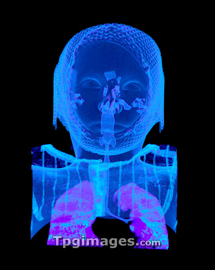
Child's head and chest, CT scan. Coloured 3-D computed tomography (CT) scan of a child's head and chest in frontal view. Within the head, the scan shows the sinuses, ear passages and nasal cavity. Within the throat and chest, the scan shows the larynx, pharynx, trachea and lungs. The scan will be used by surgeons to navigate around the brain while operating for brain cancer. This image was produced using a multi-slice CT scanner, which uses a thin X-ray beam to scan around the patient. OsiriX medical imaging software was used to reconstruct the slices into coloured 3-D images of bones and soft tissue. The program allows surgeons to navigate around the body using fly- through animations.
| px | px | dpi | = | cm | x | cm | = | MB |
Details
Creative#:
TOP03220866
Source:
達志影像
Authorization Type:
RM
Release Information:
須由TPG 完整授權
Model Release:
N/A
Property Release:
N/A
Right to Privacy:
No
Same folder images:
 Loading