
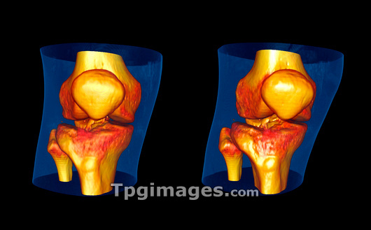
Knees, CT scan. Three-dimensional computed tomography (CT) scan of two knee joints in frontal view. The bones which comprise the knee are the distal femur, the proximal tibia and the patella (knee cap). This image was produced using a multi- slice CT scanner, which uses a thin X-ray beam to scan around the patient to create 'slices' of the body. A computer reconstructs the slices into coloured three-dimensional images including bones and soft tissue. The surgeon can navigate through the data using a 'fly-through' animation of the images. This image was created using OsiriX medical imaging software.
| px | px | dpi | = | cm | x | cm | = | MB |
Details
Creative#:
TOP03220817
Source:
達志影像
Authorization Type:
RM
Release Information:
須由TPG 完整授權
Model Release:
N/A
Property Release:
N/A
Right to Privacy:
No
Same folder images:
 Loading