
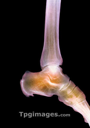
Healthy ankle. Coloured X-ray of the ankle joint of an adult woman, in side view. The bones which form the ankle joint are the shin bone (tibia, at top) with the smaller fibula (behind it). These articulate with the talus bone (uppermost foot bone) and are also attached by ligaments to the calcaneus (back of foot bone). Bones of the midfoot are also seen, with toes to the right of image. The ankle joint provides up-and-down foot movements, while tilting and rotating movements of the foot occur at joints within the foot itself.
| px | px | dpi | = | cm | x | cm | = | MB |
Details
Creative#:
TOP03220789
Source:
達志影像
Authorization Type:
RM
Release Information:
須由TPG 完整授權
Model Release:
N/A
Property Release:
N/A
Right to Privacy:
No
Same folder images:
 Loading