
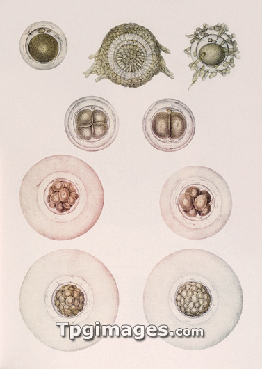
Development of an embryo, historical anatomical artwork. This 19th century textbook illustration shows the sequence of stages in embryonic development, starting at upper left. The first diagram is an ovum (egg cell) removed from the ovary of a rabbit. The second and third show the process of fertilisation with the ovum surrounded by sperm cells. Once fertilised, the cell divides (central diagrams). The final diagram shows a blastocyst, or hollow ball of cells. Development takes place in the fallopian tube, as the cells travel towards the uterus. The illustration is taken from the 19th century French textbook The Atlas of Human Anatomy and Surgery by J. M. Bourgery and N. H. Jacob.
| px | px | dpi | = | cm | x | cm | = | MB |
Details
Creative#:
TOP03220496
Source:
達志影像
Authorization Type:
RM
Release Information:
須由TPG 完整授權
Model Release:
N/A
Property Release:
N/A
Right to Privacy:
No
Same folder images:
 Loading