
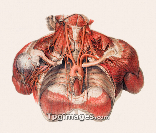
Blood vessels of the chest and neck, historical anatomical artwork. The thoracic (chest) cavity has been dissected in this anterior (front) view to reveal part of the aorta, the largest artery in the human body. The aorta forms an arch (central, u-shaped) just above the heart, and then descends through the thorax and diaphragm (bottom, dumbbell-shaped) into the abdomen. The carotid arteries emerge from the aortic arch and carry blood up the neck to the head and brain. The lungs have been removed here but the bronchi (centre, grey/white) can be seen next to the aortic arch. This illustration is taken from the 19th century French textbook The Atlas of Human Anatomy and Surgery by J. M. Bourgery and N. H. Jacob.
| px | px | dpi | = | cm | x | cm | = | MB |
Details
Creative#:
TOP03220462
Source:
達志影像
Authorization Type:
RM
Release Information:
須由TPG 完整授權
Model Release:
N/A
Property Release:
N/A
Right to Privacy:
No
Same folder images:
 Loading