
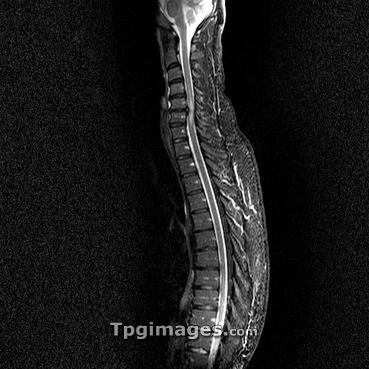
Slipped disc. Magnetic resonance imaging (MRI) scan of a sagittal (side) section through the spine of a 45 year old man showing a slipped disc (upper centre). The front of the body is at left and the spinal cord (grey) can also be seen with the brain at top. Intervertebral discs are rubbery cartilaginous pads that cushion the vertebrae (block-like bones of the spine) and give the neck and back flexibility. They can become prolapsed due to an injury or mechanical stress. Here, the slipped (prolapsed) disc between the bones of the cervical spine (neck bones C5 and C6) is compressing the spinal cord, causing swelling. This can cause pain and numbness in the region below the prolapse. Treatment is with bed rest.
| px | px | dpi | = | cm | x | cm | = | MB |
Details
Creative#:
TOP03216129
Source:
達志影像
Authorization Type:
RM
Release Information:
須由TPG 完整授權
Model Release:
N/A
Property Release:
N/A
Right to Privacy:
No
Same folder images:
 Loading