
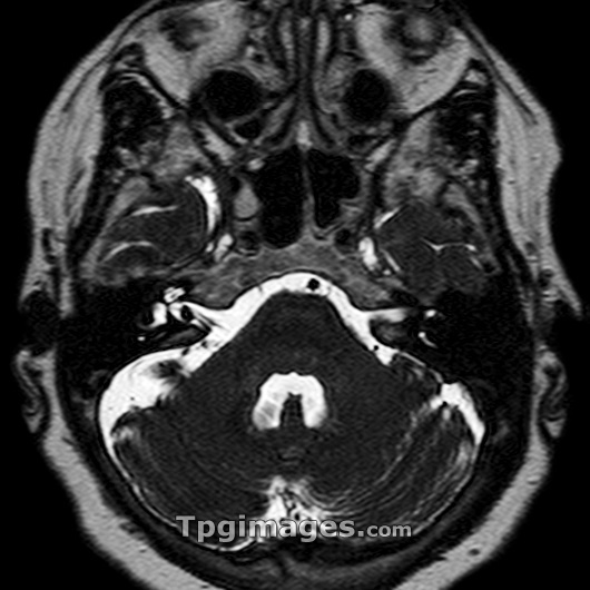
Acoustic neuroma. Magnetic resonance imaging (MRI) scan of an axial (horizontal) section through the brain of a 51 year old woman. The front of the head is at top. A 3-4 millimetre mass is visible in the internal auditory meatus (inner ear), seen as a black dot on a white background (centre left). It is most likely to represent a small acoustic neuroma (schwannoma). Acoustic neuromas are tumours of the auditory nerve of the ear, which may spread from the inner ear to the brain. They can be malignant or benign. In this case it has resulted in right sided deafness. Surgical removal is the only cure. MRI uses radio waves and a magnet to obtain inssliceins body images.
| px | px | dpi | = | cm | x | cm | = | MB |
Details
Creative#:
TOP03215478
Source:
達志影像
Authorization Type:
RM
Release Information:
須由TPG 完整授權
Model Release:
N/A
Property Release:
N/A
Right to Privacy:
No
Same folder images:
 Loading