
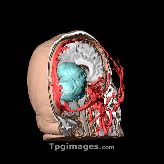
Brain tumour. 3D model of the head (rear view) of a 59-year-old woman, made up from numerous MRI (magnetic resonance imaging) scans. It is showing the internal structures of the right-hand side of the head, revealing the brain (white), and blood vessels (red). A large brain tumour, a benign (non-cancerous) meningioma, can be seen in blue at the back of the brain. Meningiomas are most often benign and arise from the meninges, the protective membranes that cover the brain and spinal cord. As it grows, a meningioma compresses adjacent brain tissue, and symptoms, such as headaches, are often related to this compression. Usually the tumour can be removed surgically, although radiotherapy may also be needed.
| px | px | dpi | = | cm | x | cm | = | MB |
Details
Creative#:
TOP03215395
Source:
達志影像
Authorization Type:
RM
Release Information:
須由TPG 完整授權
Model Release:
N/A
Property Release:
N/A
Right to Privacy:
No
Same folder images:
 Loading