
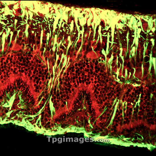
Detached retina. Confocal light micrograph of a section through a detached retina. Neurons (nerve cells) are red and glial cells (support cells) are yellow. The retina is the light-sensitive membrane that lines the back of the eyeball. If it is damaged, fluid may leak under it, causing part of it to come away from the rear wall of the eye. This retina has been detached for 4 weeks and the glial cells, which provide neurons with structural support, nutrients and oxygen, have enlarged and extended beyond their usual location of the retinal borders. This damage can cause long term complications, even after the retina is reattached. For a similar image of a healthy retina see P424/235.
| px | px | dpi | = | cm | x | cm | = | MB |
Details
Creative#:
TOP03215076
Source:
達志影像
Authorization Type:
RM
Release Information:
須由TPG 完整授權
Model Release:
N/A
Property Release:
N/A
Right to Privacy:
No
Same folder images:
 Loading