
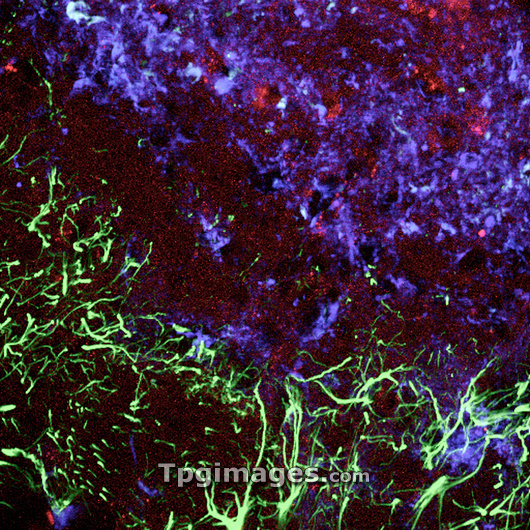
Damaged brain tissue. Confocal light micrograph of a section of damaged brain tissue. Dead neurons (nerve cells, blue) are being removed by macrophages (red) a type of white blood cell that phagocytoses (engulfs) damaged or foreign cells. The damage may lead to the formation of a scar. Enlarged glial cells (green), a type of support cell, are also seen around the damaged area. Glial cells provide structural support and oxygen and nutrients to neurons.
| px | px | dpi | = | cm | x | cm | = | MB |
Details
Creative#:
TOP03214947
Source:
達志影像
Authorization Type:
RM
Release Information:
須由TPG 完整授權
Model Release:
N/A
Property Release:
N/A
Right to Privacy:
No
Same folder images:
GLIALCELLMACROPHAGENEURONINJURYDAMAGESUPPORTCELLWHITEBLOODCELLNERVECELLBRAINENGULFINGREMOVINGSCARRINGMEDICINENEUROLOGYCONFOCALLIGHTMICROGRAPHLIGHTMICROGRAPHLIGHTMICROSCOPENEUROLOGICALMEDICALMEDICINEMEDICALHEALTHCAREDISEASEDISORDERCONDITIONDAMAGEDINJUREDDEADSCARNEURONENEURONESNEURONSCELLSMACROPHAGESENLARGEDFLUORESCENTSTAINSTAINEDIMMUNOFLUORESCENTIMMUNOFLUORESCENCEFLUORESCENCE
BLOODBRAINCELLCELLCELLCELLCELLSCONDITIONCONFOCALDAMAGEDAMAGEDDEADDISEASEDISORDERENGULFINGENLARGEDFLUORESCENCEFLUORESCENTGLIALHEALTHCAREIMMUNOFLUORESCENCEIMMUNOFLUORESCENTINJUREDINJURYLIGHTLIGHTLIGHTMACROPHAGEMACROPHAGESMEDICALMEDICALMEDICINEMEDICINEMICROGRAPHMICROGRAPHMICROSCOPENERVENEUROLOGICALNEUROLOGYNEURONNEURONENEURONESNEURONSREMOVINGSCARSCARRINGSTAINSTAINEDSUPPORTWHITE
 Loading