
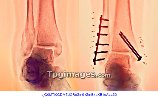
Pinned ankle fracture. Coloured frontal X-rays of the ankle bones of a patient with an ankle fracture, before (left) and after (right) the fractures were pinned. The malleolus (bony projection at the end of a bone) of both lower leg bones have been fractured and then pinned back together. The fibula (left side of each X-ray) is the thinner of the two lower leg bones, and has had a plate as well as screws used to hold it together. The pins and plates hold the bone fragments in position while they heal. The tibia (right side of each X-ray) is the thicker of the two lower leg bones. The bones at bottom are part of the ankle bones. Staples are seen in the surrounding tissue at right, showing where the skin was stapled back together following the bone- pinning operation. This is the patient's right ankle.
| px | px | dpi | = | cm | x | cm | = | MB |
Details
Creative#:
TOP01423480
Source:
達志影像
Authorization Type:
RM
Release Information:
須由TPG 完整授權
Model Release:
N/A
Property Release:
N/A
Right to Privacy:
No
Same folder images:
 Loading